CEOCFO Magazine, PO Box 340
Palm Harbor, FL 34682-0340
Phone: 727-480-7070

Email: info@ceocfocontact.com

Weekly Digital Publication IN-DEPTH INTERVIEWS WITH Top   CORPORATE EXECUTIVES (727) 480-7070 info@ceocfocontact.com FIND INTERVIEWS AND ARTICLES

Business Services | Solutions
Medical | Biotech
Cannabis  | Hemp
Banking | FinTech | Capital
Government Services
Public Companies
 Industrial | Resources

Clean Tech  
Global | Canadian


Lynn Fosse, Senior Editor

Steve Alexander, Associate Editor

Bud Wayne, Marketing
& Production Manager

Christy Rivers - Editorial Associate

INTERview


-
Print - PDF  Mobile - PDF


An interview with “The Prostate Cancer Detector”, Dr. Randall W. Jones, CEO of Bot Image, Inc.


Dr. Randall W. Jones

Founder/CEO


Bot Image, Inc.

https://www.botimageai.com/


Contact:

BOT IMAGE INC

Melanie Jones

(402) 334-2525

mjones@botimageai.com


Interview conducted by:

Lynn Fosse, Senior Editor

CEOCFO Magazine


Published – August 29, 2022


CEOCFO: Dr. Jones, what is the idea behind Bot Image?

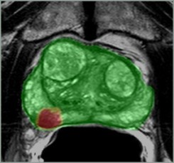
Dr. Jones: It is a dedicated artificial intelligence software company. Bot Image’s mission is to develop software tools using AI that assists radiologists and other physicians in interpreting medical diagnostic data. In the case of our first product, it is interpreting prostate MRI and helping them properly interpret or read those MRI scans.  


CEOCFO: What are some of the challenges in reading MRI scans specifically to prostate, and why is that the first area you have chosen?

Dr. Jones: Radiology of course is the offshoot of general practitioners that study how to read or interpret all of the different radiological mediums such as x-ray, CT, ultrasound, MRI, etcetera. It is fairly broad and that is why radiologists have to go beyond general physician training for a couple of years and typically perform an internship. Yet even then, most radiologists do not have a large amount of experience with any one particular diagnostic medium let alone with one particular diagnostic exam within one medium such as a prostate MRI exam. Another words, radiology is so broad and diffuse that in order to be very, very good at interpreting some medical image you have to highly specialize during a large part of your career. Here is the rest of the problem. Prostate cancer manifests itself in 1 in 8 adult males during their lifetimes and kills over 30,000 annually in the USA alone. While prostate MRI is still the best diagnostic medium for viewing this soft tissue organ and distinguishing cancerous tissues from non-cancerous (hence, specificity), the challenges of properly interpreting prostate MRI are significant, and in general, radiology performance is typically well below 85% even with the many of the experienced. Those with much less experience perform unremarkably above 50% accuracy. So, Bot Image, Inc.’s first product, ProstatIDTM is an AI algorithm that assists radiologists in better detecting and diagnosing prostate cancer.


My background is I am a PhD electrical engineer with post-doctoral experience as an MRI physicist having been a physicist for an MRI facility. I have specialized in prostate MRI combining my physics and engineering experiences, so I see this problem solution with good qualifications. I do empathize with the physician in interpreting prostate MRI because it truly is extraordinarily challenging. One must be able to accurately discern, within this soft tissue medley called a prostate; whether tissues are BPH (benign prostate hyperplasia), which is common in almost every man over 55 and worsening with age, or Prostatitis (an infection), or other things like benign cysts, all which can mimic or obscure the appearance of a cancer.


CEOCFO: What exactly is the AI looking to review?

Dr. Jones: If done properly, AI sees far more - and I use the word “sees” figuratively - than the human eye is capable of seeing.  We call these visual and non-visual clues “features”. Our particular algorithm employees 64 features of the image sets themselves as compared to the gray scale, intensity, and limited texture that the eye can discern from images. In MRI they scan with a lot of different image parameters, creating various imaging sets. These different parameters help indicate different grey scales within each set that indicate abnormalities. These various image sets are viewed by a highly trained physician or radiologists, yet the human eye, even a highly trained human eye can perceive limited features as compared to those features measured by AI; hence AI’s advantage.  


By that, if you feed an algorithm that has the ability to learn, which is what deep learning artificial intelligence is, and if you feed the algorithm with sufficient amounts of known data, in our case prostate MRI with known biopsy results, it can learn the difference between normal tissue, abnormal non-cancerous tissue and cancerous tissue. The data included large numbers of accurately localized (in 3D on the MRI data sets) biopsy data with their associated pathology lab reports. This is what researchers refer to as biological ground truth as the histopathology slide reveals cancer cellular structure to trained pathologists. In other words, we know which of those particular locations in the image are cancerous or not by the pathology, and further, we know with a high degree of accuracy that all of the rest of each image is not cancerous.   


We measure both physician and AI software accuracy in terms of sensitivity-specificity curves or receiver operating curves which measure detection ability (sensitivity) as well as the ability to differentiate between cancer and non-cancer (specificity). Again, if you were to take an average of all of prostate MRI interpreter’s performance curves in the US, you would likely be amazed that this average performance is right around 60-70 percent – this from sampling a large set of data from a large number of radiology clinics. There are exceptional radiology experts out there that can reach up close to maybe 90%, but those are exceptionally rare individuals that have had a decade or more of not only reading prostate MRI, but of getting real-time feedback from a partner urology department that has provided them feedback of how many times they got it right and how many times they got it wrong. This simulates how an artificial intelligence program ‘learns’.


Our algorithm has been trained sufficiently that it currently is operating at just a little over 90% accuracy in terms of prostate MRI detection which we believe is higher than almost all radiologists in North America. Of course, we have not and could not measure everyone but the statistical sampling as well as literature suggests this is true.  

CEOCFO: You have received FDA clearance; how did you decide when it was ready to go and what was the moment you had enough AI?

Dr. Jones: We fortunately were able to get our hands on about two thousand patient data sets. That is another discussion because it is extraordinarily challenging to get a hold of patient data because of the very strict HIPAA laws in the country. You have to find the right hospital partners who have done everything right with patient confidentiality and consent wherein you can share anonymized patient data and use it for research. So, after slicing up each prostate data set into 20-30 slices each yielded a tremendous amount of data with which to train and test the algorithm.   


So, when we achieved a 90% accuracy (0.9 sensitivity-specificity) in standalone studies of the software alone, that was the moment of a green light. This is because I knew from attending academic lectures in radiology for the past decade, that we had something that exceeded the ability of almost every radiologist on the planet. The beautiful thing was it was measured very scientifically and was not in a biased format.  We had set aside the appropriate number of studies and did not train on those studies; hence, the algorithm saw these studies for the first time during its test. That is just good practice in terms of software development. When we achieved those results, I knew we were ready to go.


CEOCFO: Why are we going to need radiologists in the future?

Dr. Jones: Artificial intelligence does not do it all even if it is 90% accurate; or even if it approaches 100% - which I believe it will given enough data and tweaking of the algorithm. There are those times when there is some other anomaly that it may not have seen, and there are other underlying conditions that need to be taken into consideration in terms of that patient diagnosis and/or treatment. The radiologic exam is just the first part of this patient’s path through the medical system. The radiologist is important to understand and interpret what the AI has said, agree with it or not, and then put that in their report, which of course is basically a recommendation for the primary care physician to consider in terms of treatment planning or where they go next. Also, other factors such as sexual activity, age, family history - all these things need to be considered in terms of what is the best outcome for this particular patient. So, I don’t think that radiologists are ever going to be obsoleted – at least not those that accept and adapt AI. I like to look at it like this. Creators of diagnostic AI are making products to improve diagnostic performance while making them faster and more accurate; thus improving patient outcome. It is a win/win for everybody. What is going to happen, or should happen, is that the price should come down but the volume should go up significantly.


One of my dreams and goals, was and remains, to improve healthcare for men just like the big push was for women over a decade ago in terms of breast cancer. We are way behind with prostate cancer, way behind, but slowly catching up. Today there are still probably hundreds of thousands if not millions of prostatectomies being done when they should not have been done. Could you imagine if you have a lump in your breast as a female and that lump turns out to be a cancer, do you want to go have a mastectomy, or maybe just have technology go in and remove that cancerous legion and keep your breast? I think that is a pretty easy choice; yet it has only recently started to be a choice provided to men with prostate cancer where some type of focal lesion ablation has been approved by insurance.


CEOCFO: Why has the prostate been treated like a red-headed stepchild?

Dr. Jones: I think it is just publicity and public awareness. This could be interpreted as a sexist conversation but I don’t intend for it to be. The female breast has always gotten and continues to get a lot of attention in advertisement, entertainment/movies, modeling, and self-image. Breast cancer was too prevalent, as were mastectomies, and causing grief to all affected; hence, it was society who made the decision to say ‘let’s just go focus on breast cancer and improve this standard of care’. And the medical community did just that. I don’t pretend to know the answer to your question, but I feel that the same kind of movement has begun to catch-on regarding prostate cancer.  


Prostate cancer awareness is gaining more steam and momentum. We have prostate awareness month and many non-profit companies focused on this problem and solutions while providing education to society. My frustration is that the medical community is still very slow to adopt to change despite significant peer-reviewed evidence, but guess what is even slower to change? The insurance industry. This industry dictates and restricts what the medical community can do, and from my extensive experience at introducing new medical devices and processes, they are typically 10 years behind where research is. Unfortunately then, physicians often cannot employ what they know is best because of this delayed approval process. This topic is near and dear to me and I discuss it further in my recently published book (ForbesBooks) titled “The Healthcare Disruptor”.


CEOCFO: How are you commercializing; what is your plan for Bot Image?

Dr. Jones: We have already created a SaaS, Software as a Service, product out of this which requires less than an hour time on the part of the hospital or imaging facility IP personnel to connect and become operational. We simply send them an IT setup sheet/guide. They fill it out, they can get on the phone with our IT department and ping each other to confirm we have communications, and then they are set and ready to go. We have been running this in beta test sites for over a year and have also started to make our first sales and contracts of volume prostate interpretation with facilities.


The way this works is the radiology department connects as above and they merely “push” the MRI data to us, we process it, and they get the results back in five minutes or less - real-time diagnostics and detection. The doctor can still have the patient in the MRI bore being further scanned and he/she will get results back soon after they send us the three requisite scanning series of that patient study. So we are already in business with this SaaS product. We also have a working model of software that can exist behind the hospital firewall. A lot of the times academic institutions just simply will not let the data be pushed out even though it is a cyber-secure, HIPAA compliant communication, similar to a remote radiologist would have in his home, so this version of the software abates that issue entirely. We also have other R&D in-works on additional AI products to be discussed as we near completion.


Bot Image continues to break down the walls of resistance in terms of accepting new technology which takes good marketing and academic studies. We have been busy educating the radiological community about the existence of ProstatID through academic partnerships and publications. We had one academic paper accepted and I presented it at the International Society of Magnetic Resonance in Medicine (ISMRM) in London this spring. We have three other academic papers that have been submitted from three different academic partners who experienced using the software. Once the academic papers are published/presented, we expect to see the early adopters of this technology then followed by the masses. We are already seeing that from our own website and our own marketing and advertisement. It is beginning to turn that really large slow-moving ship that is the medical community. Our academic partners are helping us state that we have a tool that is really beneficial here that is going to help them, and more importantly, mankind.


Bot Image, Inc. | Dr. Randall W. Jones | Prostate Cancer Detection | Prostate Cancer Artificial Intelligence | AI for Prostate Cancer | AI for Radiologists | QT Prostate MRI | ProstatID | An interview with “The Prostate Cancer Detector”, Dr. Randall W. Jones, CEO of Bot Image, Inc. | CEO Interviews 2022 | Medical Companies | Artificial Intelligence for MRI, prostate Cancer detection methods, prostate cancer detection test, ProstatIDTM, artificial intelligence software for MRI system, prostate cancer detection using AI and MRI, prostate cancer AI, prostate MRI AI, prostate AI MRI, MRI AI, AI MRI, medical AI, bot image, bot image AI, artificial intelligence, prostate artificial intelligence, artificial intelligence for prostate cancer, artificial intelligence for MRI, Bot Image, Inc. Press Releases, News, Linkedin, Facebook, Twitter

“One must be able to accurately discern, within this soft tissue medley called a prostate; whether tissues are BPH (benign prostate hyperplasia), which is common in almost every man over 55 and worsening with age, or Prostatitis (an infection), or other things like benign cysts, all which can mimic or obscure the appearance of a cancer.”
Dr. Randall W. Jones


HOME

CURRENT ISSUE

INTERVIEW INDEX

CEOCFO SERVICES

CEOCFO MOBILE


“Bot Image’s mission is to develop software tools using AI that assists radiologists and other physicians in interpreting medical diagnostic data.”
Dr. Randall W. Jones

“Our algorithm has been trained sufficiently that it currently is operating at just a little over 90% accuracy in terms of prostate MRI detection which we believe is higher than almost all radiologists in North America.”
Dr. Randall W. Jones


Latest News

“Bot Image, Inc. receives FDA clearance for ProstatIDTM, an AI software to assist radiologists in detection and diagnosis of Prostate cancer using MRI”